Case Report Archives - Critical Care Science (CCS)

Abstract
Rev Bras Ter Intensiva. 2021;33(3):457-460
DOI 10.5935/0103-507X.20210067
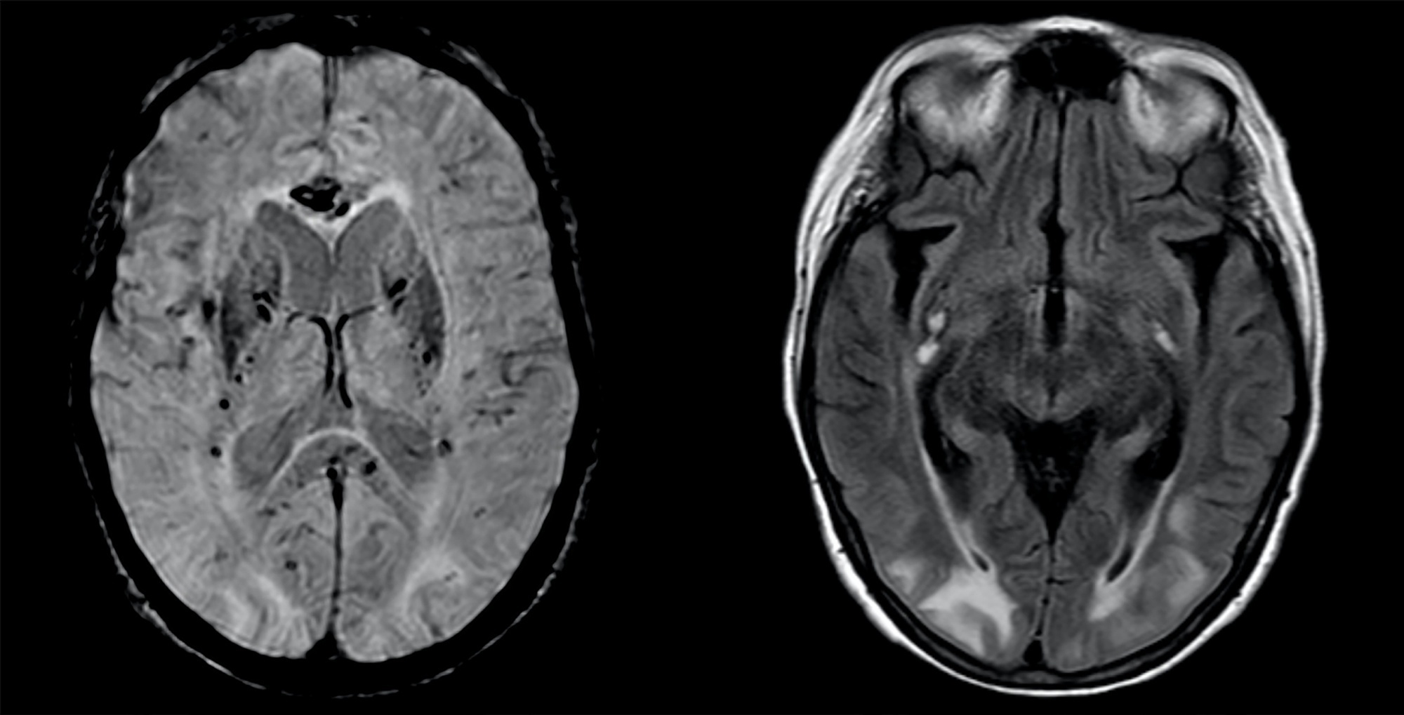
A 63-year-old woman presented to the emergency department with an acute history of fever, prostration and dyspnea. She was diagnosed with severe COVID-19 acute respiratory distress syndrome and, despite optimized critical care support, met the indications for veno-venous extracorporeal membrane oxygenation. On day 34, after 7 days of wean sedation with a positive evolution of neurologic status, she presented a limited generalized tonic-clonic seizure not related to hydroelectrolytic or metabolic imbalance, which led to a diagnostic investigation; her brain imaging tests showed a posterior reversible encephalopathy syndrome. This case emphasizes the issue of neurological complications in patients with severe COVID-19 infection and the importance of early diagnosis and support.
Abstract
Rev Bras Ter Intensiva. 2021;33(3):457-460
DOI 10.5935/0103-507X.20210067
A 63-year-old woman presented to the emergency department with an acute history of fever, prostration and dyspnea. She was diagnosed with severe COVID-19 acute respiratory distress syndrome and, despite optimized critical care support, met the indications for veno-venous extracorporeal membrane oxygenation. On day 34, after 7 days of wean sedation with a positive evolution of neurologic status, she presented a limited generalized tonic-clonic seizure not related to hydroelectrolytic or metabolic imbalance, which led to a diagnostic investigation; her brain imaging tests showed a posterior reversible encephalopathy syndrome. This case emphasizes the issue of neurological complications in patients with severe COVID-19 infection and the importance of early diagnosis and support.
Abstract
Rev Bras Ter Intensiva. 2021;33(2):331-335
DOI 10.5935/0103-507X.20210043
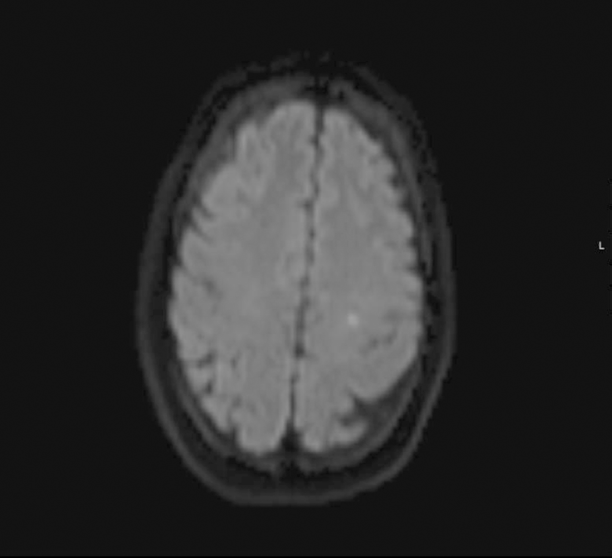
A 37-year-old woman (35 weeks pregnant) was admitted to a local hospital due to severe epistaxis resulting in shock and the need for emergency cesarean section. After failure to tamponade the bleeding, angiographic treatment was provided. After the procedure, she was admitted to the neurocritical intensive care unit and was confused and agitated, requiring sedation and endotracheal intubation. In the intensive care unit, diagnostic investigations included brain magnetic resonance imaging, lumbar puncture with viral panel, electroencephalogram, tests for autoimmunity, and hydroelectrolytic and metabolic evaluations. Magnetic resonance imaging showed a puntiform restricted diffusion area on the left corona radiata on diffusion weighted imaging and mild cortical posterior edema (without restricted diffusion), and an electroencephalogram showed moderate diffuse slow activity and fronto-temporal slow activity of the left hemisphere with associated scarce paroxysmal components. The other exams did not show any relevant alterations. Due to the temporal relationship, the clinical history and the magnetic resonance imaging results, a diagnosis of contrast-induced encephalopathy was made. After 2 days in the intensive care unit, sedation was withdrawn, the patient was extubated, and total neurological recovery was verified within the next 24 hours.
Abstract
Rev Bras Ter Intensiva. 2021;33(2):331-335
DOI 10.5935/0103-507X.20210043
A 37-year-old woman (35 weeks pregnant) was admitted to a local hospital due to severe epistaxis resulting in shock and the need for emergency cesarean section. After failure to tamponade the bleeding, angiographic treatment was provided. After the procedure, she was admitted to the neurocritical intensive care unit and was confused and agitated, requiring sedation and endotracheal intubation. In the intensive care unit, diagnostic investigations included brain magnetic resonance imaging, lumbar puncture with viral panel, electroencephalogram, tests for autoimmunity, and hydroelectrolytic and metabolic evaluations. Magnetic resonance imaging showed a puntiform restricted diffusion area on the left corona radiata on diffusion weighted imaging and mild cortical posterior edema (without restricted diffusion), and an electroencephalogram showed moderate diffuse slow activity and fronto-temporal slow activity of the left hemisphere with associated scarce paroxysmal components. The other exams did not show any relevant alterations. Due to the temporal relationship, the clinical history and the magnetic resonance imaging results, a diagnosis of contrast-induced encephalopathy was made. After 2 days in the intensive care unit, sedation was withdrawn, the patient was extubated, and total neurological recovery was verified within the next 24 hours.
Abstract
Rev Bras Ter Intensiva. 2021;33(2):325-325
DOI 10.5935/0103-507X.20210042
COVID-19 was declared a pandemic by the World Health Organization on March 11, 2020. The clinical presentation is predominantly respiratory symptoms; however, in the current literature, several neurological manifestations associated with SARS-CoV-2 infection have been described. The authors present the clinical case of a 45-year-old man hospitalized for pneumonia with a positive test result for SARS-CoV-2, without a neurological history, who, on the sixteenth day of hospitalization, presented a sudden change in his state of consciousness accompanied by conjugated right gaze deviation and myoclonus of the face and thoracic region to the left, followed by generalized tonic-clonic seizures associated with persistent left hemiparesis. The present study highlights a positive RT-PCR test for SARS-CoV-2 in cerebrospinal fluid. The patient progressed with gradual improvement, and the outcome was favorable.
Abstract
Rev Bras Ter Intensiva. 2021;33(2):320-324
DOI 10.5935/0103-507X.20210041
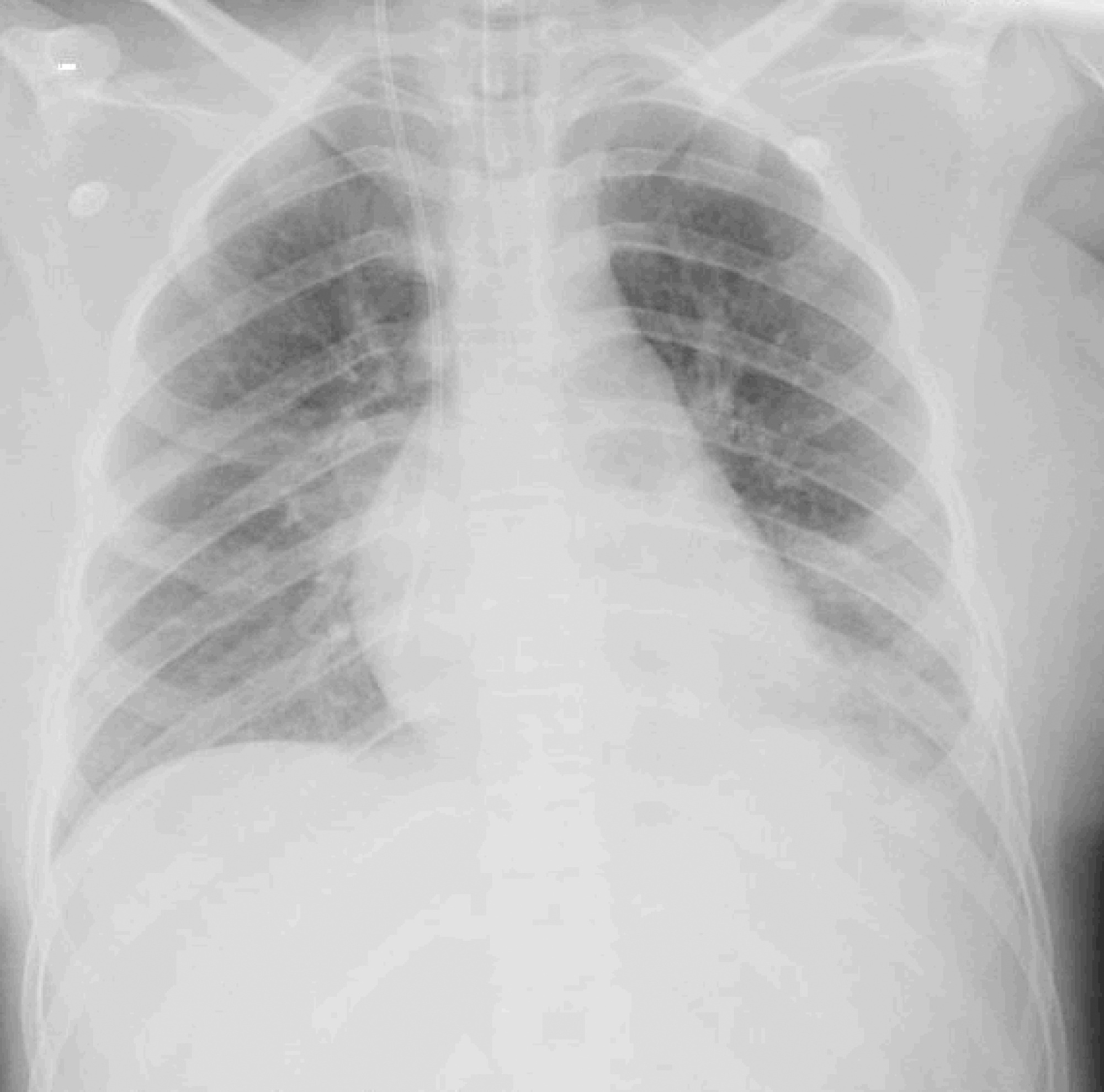
We report a case of Influenza B infection and Kawasaki disease in an adolescent, diagnosed during the COVID-19 pandemic. An asthmatic female adolescent presented with fever and flu-like symptoms for 7 days and was admitted with acute respiratory failure requiring mechanical ventilation. She progressed with hemodynamic instability responsive to vasoactive drugs. Antibiotic therapy and support measures were introduced, showing progressive hemodynamics and respiratory improvement, however with persistent fever and increased inflammatory markers. During the hospitalization, she developed bilateral non-purulent conjunctivitis, hand and feet desquamation, strawberry tongue, and cervical adenopathy, and was diagnosed with Kawasaki disease. She was prescribed intravenous immunoglobulin and, due to the refractory clinical conditions, corticosteroid therapy was added; 24 hours later, the patient was afebrile. No coronary changes were found. A full viral panel including COVID-19 C-reactive protein and serology could only isolate the Influenza B virus. During the hospitalization, she was diagnosed with pulmonary thromboembolism; coagulopathies were investigated, and she was diagnosed with heterozygous factor V Leiden mutation. There is a potential association between Kawasaki disease and infection with Influenza B or with other viruses such as coronavirus. Therefore, this association should be considered in pediatric patients, adolescents included, with prolonged febrile conditions.
Abstract
Rev Bras Ter Intensiva. 2021;33(2):320-324
DOI 10.5935/0103-507X.20210041
We report a case of Influenza B infection and Kawasaki disease in an adolescent, diagnosed during the COVID-19 pandemic. An asthmatic female adolescent presented with fever and flu-like symptoms for 7 days and was admitted with acute respiratory failure requiring mechanical ventilation. She progressed with hemodynamic instability responsive to vasoactive drugs. Antibiotic therapy and support measures were introduced, showing progressive hemodynamics and respiratory improvement, however with persistent fever and increased inflammatory markers. During the hospitalization, she developed bilateral non-purulent conjunctivitis, hand and feet desquamation, strawberry tongue, and cervical adenopathy, and was diagnosed with Kawasaki disease. She was prescribed intravenous immunoglobulin and, due to the refractory clinical conditions, corticosteroid therapy was added; 24 hours later, the patient was afebrile. No coronary changes were found. A full viral panel including COVID-19 C-reactive protein and serology could only isolate the Influenza B virus. During the hospitalization, she was diagnosed with pulmonary thromboembolism; coagulopathies were investigated, and she was diagnosed with heterozygous factor V Leiden mutation. There is a potential association between Kawasaki disease and infection with Influenza B or with other viruses such as coronavirus. Therefore, this association should be considered in pediatric patients, adolescents included, with prolonged febrile conditions.
Abstract
Rev Bras Ter Intensiva. 2021;33(1):167-171
DOI 10.5935/0103-507X.20210018
The natural history of the disease, and the treatment of post-COVID-19 patients, are still being built. Symptoms are persistent, even in mild cases, and the infection consequences include fatigue, dyspnea, tachycardia, muscle loss, and reduced functional capacity. Regarding cardiopulmonary rehabilitation, there seems to be an improvement in functional capacity, quality of life, and prognosis with the 6-Minute Walk Test used as a prognostic and therapeutic evaluator. Therefore, this case series report aims to present our experience with four cases of different severity levels, involved in a post-COVID-19 cardiopulmonary rehabilitation program. These patients were assessed with the 6-Minute Walk Test, peripheral muscle strength, and double product at rest, to assess the results after a three-month rehabilitation protocol of at least 300 minutes per week. The four patients had their distance covered during the walk test increased between 16% and 94%. Peripheral muscle strength was improved by 20% to six times the baseline values, and double product at rest was reduced by 8% to 42%. The cardiopulmonary rehabilitation program had a positive impact on these cases, improving functional capacity despite the different severity levels in these post-COVID-19 cases.
Abstract
Rev Bras Ter Intensiva. 2021;33(1):172-175
DOI 10.5935/0103-507X.20210019
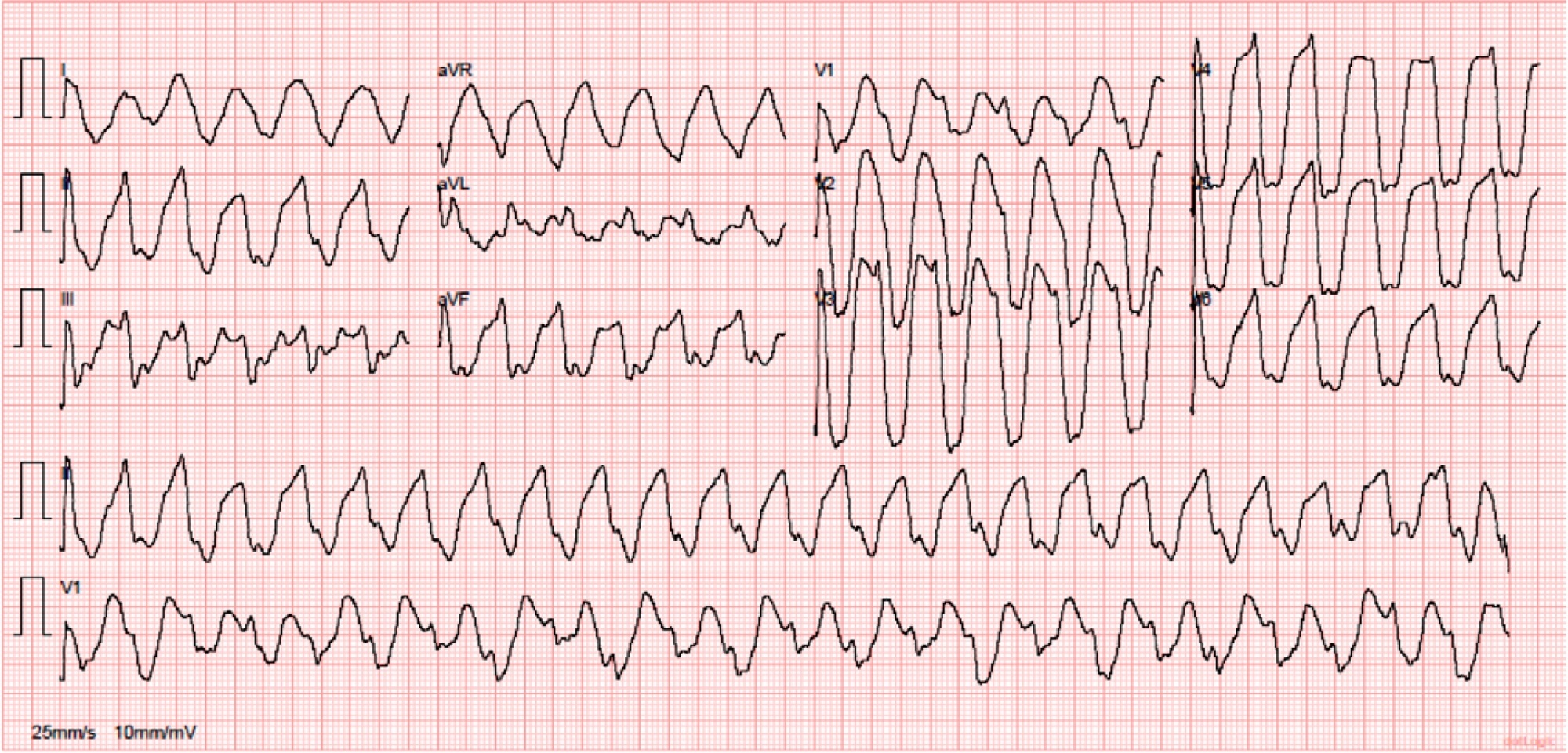
European yew (Taxus baccata) is a tree with alternate branchlets, green needles and reddish-brown bark. A high-dose ingestion of Taxus baccata for suicidal purposes usually results in death. The systemic toxicity is mainly cardiac. The authors describe the case of a young patient who ingested a high dose of yew needles and presented to the emergency department with a serious intoxication, which manifested as a chaotic malignant arrhythmia that was successfully treated after exhaustive supportive care.
Abstract
Rev Bras Ter Intensiva. 2021;33(1):172-175
DOI 10.5935/0103-507X.20210019
European yew (Taxus baccata) is a tree with alternate branchlets, green needles and reddish-brown bark. A high-dose ingestion of Taxus baccata for suicidal purposes usually results in death. The systemic toxicity is mainly cardiac. The authors describe the case of a young patient who ingested a high dose of yew needles and presented to the emergency department with a serious intoxication, which manifested as a chaotic malignant arrhythmia that was successfully treated after exhaustive supportive care.
Abstract
Rev Bras Ter Intensiva. 2020;32(4):606-610
DOI 10.5935/0103-507X.20200099
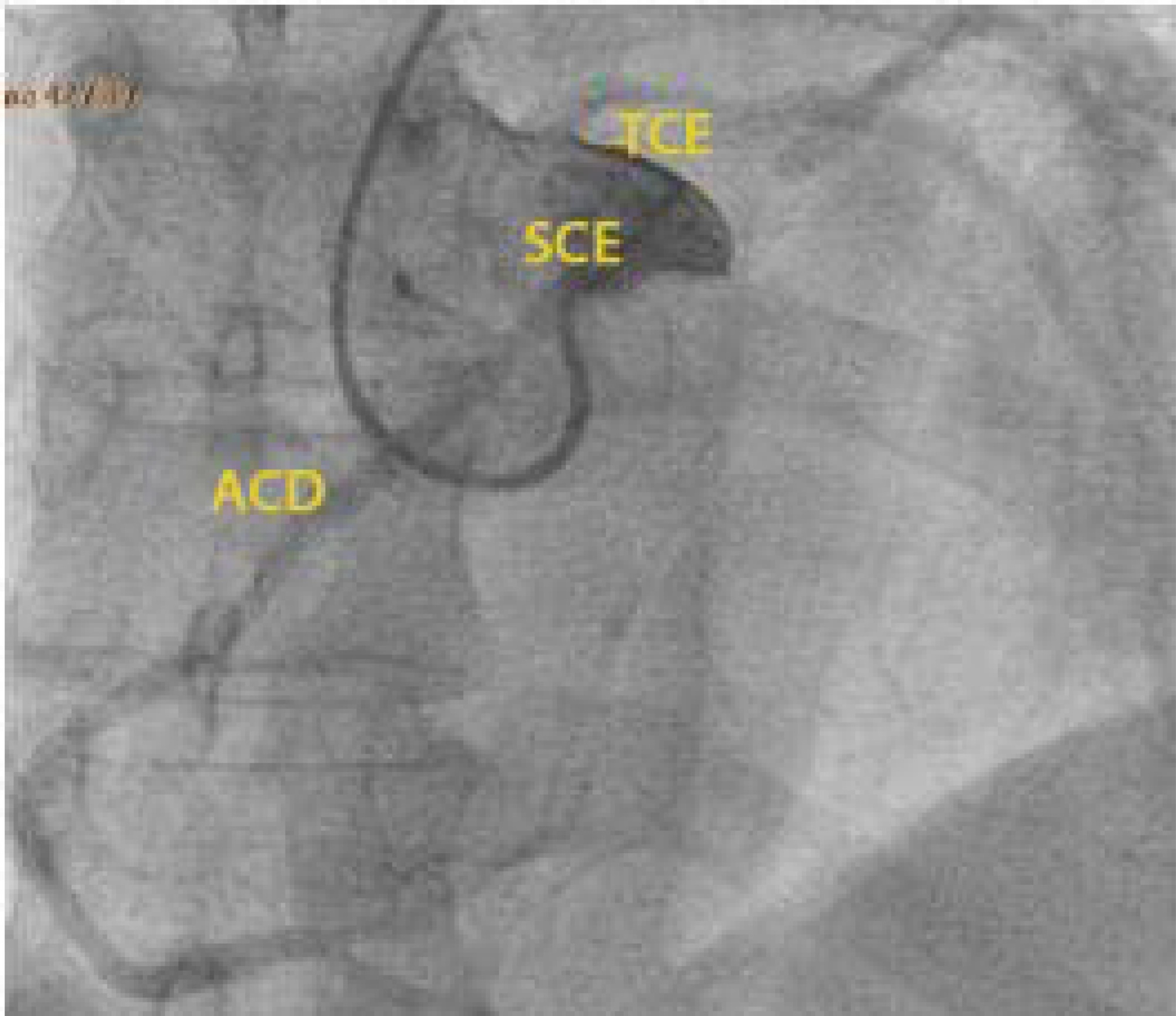
The authors report a rare case of successful Advanced Life Support in the context of cardiac arrest due to the presence of an anomalous aortic origin of the right coronary artery in a 49-year-old patient. The patient was admitted due to chest pain and dyspnea, with rapid evolution of pulseless ventricular tachycardia and cardiopulmonary arrest. Acute myocardial infarction was considered, and in the absence of a hemodynamic laboratory in the hospital, thrombolysis was performed. Subsequently, coronary angiography revealed no angiographic lesions in the coronary arteries and an anomalous right coronary artery originating from the opposite sinus of Valsalva. Coronary computed tomography angiography confirmed this finding and determined the course between the pulmonary artery and the aorta. The patient underwent cardiac surgery with a bypass graft to the right coronary artery, with no recurrent episodes of arrythmia.
Abstract
Rev Bras Ter Intensiva. 2020;32(4):606-610
DOI 10.5935/0103-507X.20200099
The authors report a rare case of successful Advanced Life Support in the context of cardiac arrest due to the presence of an anomalous aortic origin of the right coronary artery in a 49-year-old patient. The patient was admitted due to chest pain and dyspnea, with rapid evolution of pulseless ventricular tachycardia and cardiopulmonary arrest. Acute myocardial infarction was considered, and in the absence of a hemodynamic laboratory in the hospital, thrombolysis was performed. Subsequently, coronary angiography revealed no angiographic lesions in the coronary arteries and an anomalous right coronary artery originating from the opposite sinus of Valsalva. Coronary computed tomography angiography confirmed this finding and determined the course between the pulmonary artery and the aorta. The patient underwent cardiac surgery with a bypass graft to the right coronary artery, with no recurrent episodes of arrythmia.
Abstract
Rev Bras Ter Intensiva. 2020;32(4):603-605
DOI 10.5935/0103-507X.20200098
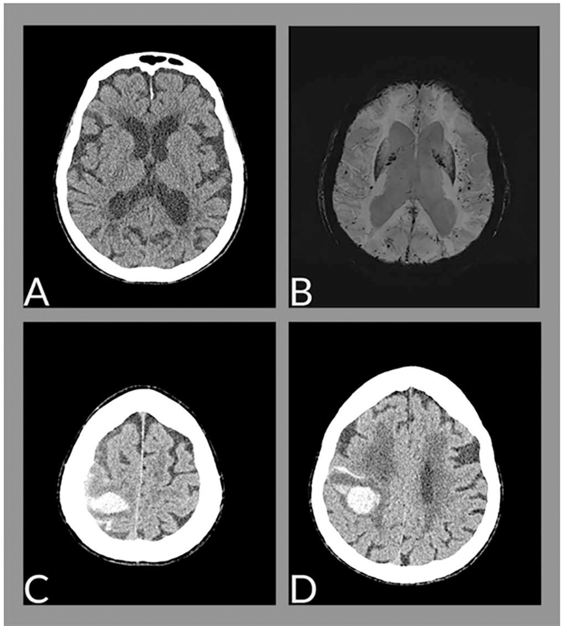
The neurological changes associated with COVID-19 have been frequently described, especially in cases of greater severity, and are related to multifactorial causes, such as endothelial dysfunction, inflammatory mediator release (cytokine storm), endothelial dysfunction and hypoxemia. We report the case of a female patient, 88 years old, with cerebral hemorrhage associated with amyloid angiopathy in the context of SARS-CoV-2 infection.
Abstract
Rev Bras Ter Intensiva. 2020;32(4):603-605
DOI 10.5935/0103-507X.20200098
The neurological changes associated with COVID-19 have been frequently described, especially in cases of greater severity, and are related to multifactorial causes, such as endothelial dysfunction, inflammatory mediator release (cytokine storm), endothelial dysfunction and hypoxemia. We report the case of a female patient, 88 years old, with cerebral hemorrhage associated with amyloid angiopathy in the context of SARS-CoV-2 infection.