The neurological changes associated with COVID-19 have been frequently described, especially in cases of greater severity, and are related to multifactorial causes, such as endothelial dysfunction, inflammatory mediator release (cytokine storm), endothelial dysfunction and hypoxemia. We report the case of a female patient, 88 years old, with cerebral hemorrhage associated with amyloid angiopathy in the context of SARS-CoV-2 infection.
Ordinola AAM, Osmar SS, Marussi VHR, Rojas SSO, Baeta AM, Chaddad Neto FE, Veiga VC. Cerebral hemorrhage during the active phase of SARS-CoV-2 infection in a patient with amyloid angiopathy: case report. Rev Bras Ter Intensiva 2021;32(4):603-5.
Electronic Document Format (ABNT)
Ordinola, Amanda Ayako Minemura; Osmar, Samir Sari; Marussi, Victor Hugo Rocha; Rojas, Salomón Soriano Ordinola; Baeta, Alex Machado; Chaddad Neto, Feres Eduardo; Veiga, Viviane Cordeiro. Cerebral hemorrhage during the active phase of SARS-CoV-2 infection in a patient with amyloid angiopathy: case report. Rev Bras Ter Intensiva, v. 32, n. 4, p. 603-605, Jan. 2021.
Electronic Document Format (APA)
Ordinola, A. A. M., Osmar, S. S., Marussi, V. H. R., Rojas, S. S. O., Baeta, A. M., Chaddad Neto, F. E., & Veiga, V. C. (2021). Cerebral hemorrhage during the active phase of SARS-CoV-2 infection in a patient with amyloid angiopathy: case report. Rev Bras Ter Intensiva, 32(4), 603-605.
Electronic Document Format (ISO)
Ordinola, Amanda Ayako Minemura and Osmar, Samir Sari and Marussi, Victor Hugo Rocha and Rojas, Salomón Soriano Ordinola and Baeta, Alex Machado and Chaddad Neto, Feres Eduardo and Veiga, Viviane Cordeiro. Cerebral hemorrhage during the active phase of SARS-CoV-2 infection in a patient with amyloid angiopathy: case report. Rev Bras Ter Intensiva [online]. 2021, vol. 32, n. 4, [cited 2024-05-20], pp.603-605. Available from: <https://criticalcarescience.org/article/cerebral-hemorrhage-during-the-active-phase-of-sars-cov-2-infection-in-a-patient-with-amyloid-angiopathy-case-report/>.
✕
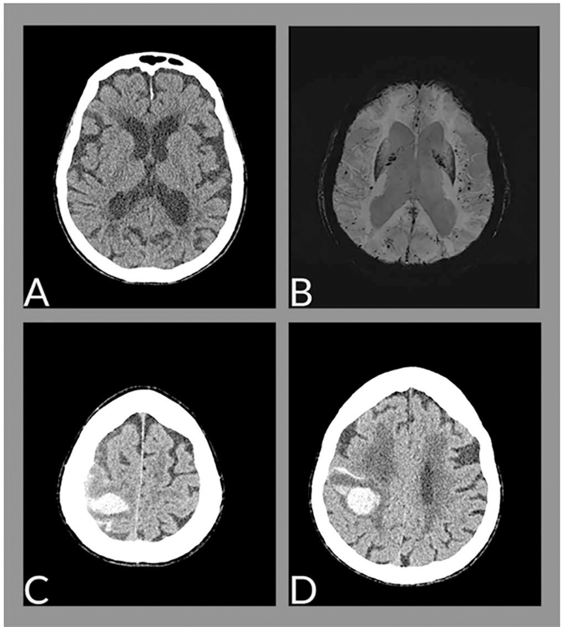
Figura 1 | (A) Imagem axial de tomografia de crânio, na admissão da paciente no hospital, mostrando alterações senis sem evidência de hemorragia. (B) Imagem axial de ressonância, sequência SWI minIP (imagem ponderada em suscetibilidade) demonstra múltiplos focos de depósito de hemossiderina com predomínio na periferia do parênquima cerebral (padrão sugestivo de angiopatia amiloide). (C e D) Imagens axiais de tomografia, demonstram hemorragias intraparenquimatosa e subaracnoide na transição frontoparietal direita.


Comments