-
Fulfilled promises to enter into a new era!
Rev Bras Ter Intensiva. 2007;19(4):413-413
-
The prevalence of nosocomial infection in Intensive Care Units in the State of Rio Grande do Sul
Rev Bras Ter Intensiva. 2007;19(4):414-420
Abstract
The prevalence of nosocomial infection in Intensive Care Units in the State of Rio Grande do Sul
Rev Bras Ter Intensiva. 2007;19(4):414-420
DOI 10.1590/S0103-507X2007000400002
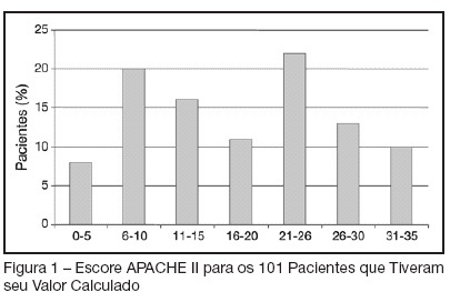
Views0See moreBACKGROUND AND OBJECTIVES: To determine the prevalence of intensive care unit (ICU)-acquired infections and the risk factors for these infections, identify the predominant infecting organisms, and evaluate the relationship between ICU-acquired infection and mortality. METHODS: A 1-day point prevalence study. Sixteen ICU of the State of Rio Grande do Sul-Brazil, excluding coronary care and pediatric units. All patients < 12 yrs occupying an ICU bed over a 24-hour period. The 16 ICU provided 174 case reports. Main outcomes: rates of ICU-acquired infection, resistance patterns of microbiological isolates, and potential risks factors for ICU-acquired infection and death. RESULTS: A total of 122 patients (71%) was infected and 51 (29%) had ICU-acquired infection. Pneumonia (58.2%), lower tract respiratory infection (22.9%), urinary tract infection (18%) were the most frequents types of ICU infection. Most frequently microorganisms reported were staphylococcus aureus (42% [64% resistant to oxacilin]) and pseudomonas aeruginosa (31%). Six risk factors for ICU acquired infection were identified: urinary catheterization, central vascular line, tracheal intubation for prolonged time (> 4 days), chronic disease and increased length of ICU stay (> 30 days). The risks factors associated with death were age, APACHE II, organ dysfunction, and tracheal intubation with or without mechanical ventilation. CONCLUSIONS: ICU-acquired infection is common and often associated with microbiological isolates of resistant organisms. This study may serve as an epidemiological reference to help the discussion of regional infection control policies.
Views0

Abstract
The prevalence of nosocomial infection in Intensive Care Units in the State of Rio Grande do Sul
Rev Bras Ter Intensiva. 2007;19(4):414-420
DOI 10.1590/S0103-507X2007000400002
Views0See moreBACKGROUND AND OBJECTIVES: To determine the prevalence of intensive care unit (ICU)-acquired infections and the risk factors for these infections, identify the predominant infecting organisms, and evaluate the relationship between ICU-acquired infection and mortality. METHODS: A 1-day point prevalence study. Sixteen ICU of the State of Rio Grande do Sul-Brazil, excluding coronary care and pediatric units. All patients < 12 yrs occupying an ICU bed over a 24-hour period. The 16 ICU provided 174 case reports. Main outcomes: rates of ICU-acquired infection, resistance patterns of microbiological isolates, and potential risks factors for ICU-acquired infection and death. RESULTS: A total of 122 patients (71%) was infected and 51 (29%) had ICU-acquired infection. Pneumonia (58.2%), lower tract respiratory infection (22.9%), urinary tract infection (18%) were the most frequents types of ICU infection. Most frequently microorganisms reported were staphylococcus aureus (42% [64% resistant to oxacilin]) and pseudomonas aeruginosa (31%). Six risk factors for ICU acquired infection were identified: urinary catheterization, central vascular line, tracheal intubation for prolonged time (> 4 days), chronic disease and increased length of ICU stay (> 30 days). The risks factors associated with death were age, APACHE II, organ dysfunction, and tracheal intubation with or without mechanical ventilation. CONCLUSIONS: ICU-acquired infection is common and often associated with microbiological isolates of resistant organisms. This study may serve as an epidemiological reference to help the discussion of regional infection control policies.
-
Pseudomonas aeruginosa: frequency of resistance to multiple drugs and cross-resistance between antimicrobials in Recife/PE
Rev Bras Ter Intensiva. 2007;19(4):421-427
Abstract
Pseudomonas aeruginosa: frequency of resistance to multiple drugs and cross-resistance between antimicrobials in Recife/PE
Rev Bras Ter Intensiva. 2007;19(4):421-427
DOI 10.1590/S0103-507X2007000400003
Views0See moreBACKGROUND AND OBJECTIVES: The frequency of multiple-antibiotic resistant bacteria has been increasing in recent years. Among the gram-negative bacteria Pseudomonas aeruginosa (P. aeruginosa) shows a great propensity for the development of multidrug resistance mechanisms. The objective of this study was to identify the profile of susceptibility to antibiotics, the frequency of multidrug resistance and the cross-resistance between drugs of P. aeruginosa strains in two tertiary hospitals in Recife, Pernambuco. METHODS: The study was carried out between September 2004 and January 2006. The antimicrobial susceptibility testing was performed in 304 strains of P. aeruginosa by the disc diffusion method in accordance with National Committee for Clinical and Laboratory Standards (NCCLS) guidelines. RESULTS: The most frequent materials were urine (26.7%) and respiratory tract secretion (26.1%) The antibiotics tested and their respective susceptibilities were as follows: piperacillin-tazobactam (66.2%); aztreonam (59.8%); amikacin (59.4%); meropenem (58.2%); imipenem (57.7%); ciprofloxacin (49.7%); gentamicin and cefepime (48.6%); ceftazidime (30%) and cefotaxime (6.8%). A high prevalence of multi-resistance was detected. Half (49.7%) the strains showed resistance to three or more antibiotics and 28% were resistant to six antimicrobials or more. Also, cross-resistance between the beta-lactams (carbapenems and piperacilin/tazobactam) and aminoglicosides and quinolones was between 22.9% and 38.1%. These drugs are commonly combined in the treatment of severe infections caused by Pseudomonas, which reflects the difficulty in choosing the appropriate option for combination therapy. CONCLUSIONS: The frequency of multidrug-resistant strains of P. aeruginosa in this study was similar to other hospitals in Brazil and higher than in other countries. In order to reduce the frequency of these multiresistant clones, epidemiologic surveillance and the rational use of antibiotic protocols need to be urgently implemented.
Views0Abstract
Pseudomonas aeruginosa: frequency of resistance to multiple drugs and cross-resistance between antimicrobials in Recife/PE
Rev Bras Ter Intensiva. 2007;19(4):421-427
DOI 10.1590/S0103-507X2007000400003
Views0See moreBACKGROUND AND OBJECTIVES: The frequency of multiple-antibiotic resistant bacteria has been increasing in recent years. Among the gram-negative bacteria Pseudomonas aeruginosa (P. aeruginosa) shows a great propensity for the development of multidrug resistance mechanisms. The objective of this study was to identify the profile of susceptibility to antibiotics, the frequency of multidrug resistance and the cross-resistance between drugs of P. aeruginosa strains in two tertiary hospitals in Recife, Pernambuco. METHODS: The study was carried out between September 2004 and January 2006. The antimicrobial susceptibility testing was performed in 304 strains of P. aeruginosa by the disc diffusion method in accordance with National Committee for Clinical and Laboratory Standards (NCCLS) guidelines. RESULTS: The most frequent materials were urine (26.7%) and respiratory tract secretion (26.1%) The antibiotics tested and their respective susceptibilities were as follows: piperacillin-tazobactam (66.2%); aztreonam (59.8%); amikacin (59.4%); meropenem (58.2%); imipenem (57.7%); ciprofloxacin (49.7%); gentamicin and cefepime (48.6%); ceftazidime (30%) and cefotaxime (6.8%). A high prevalence of multi-resistance was detected. Half (49.7%) the strains showed resistance to three or more antibiotics and 28% were resistant to six antimicrobials or more. Also, cross-resistance between the beta-lactams (carbapenems and piperacilin/tazobactam) and aminoglicosides and quinolones was between 22.9% and 38.1%. These drugs are commonly combined in the treatment of severe infections caused by Pseudomonas, which reflects the difficulty in choosing the appropriate option for combination therapy. CONCLUSIONS: The frequency of multidrug-resistant strains of P. aeruginosa in this study was similar to other hospitals in Brazil and higher than in other countries. In order to reduce the frequency of these multiresistant clones, epidemiologic surveillance and the rational use of antibiotic protocols need to be urgently implemented.
-
Presence of respiratory pathogens in the oral biofilm of patients with nosocomial pneumonia
Rev Bras Ter Intensiva. 2007;19(4):428-433
Abstract
Presence of respiratory pathogens in the oral biofilm of patients with nosocomial pneumonia
Rev Bras Ter Intensiva. 2007;19(4):428-433
DOI 10.1590/S0103-507X2007000400004
Views0See moreBACKGROUND AND OBJECTIVES: Hospitalized patients receiving treatment at intensive care units (ICU) usually show poor oral hygiene, and may have the mouth and oropharingeal region colonized by pathogens involved in nosocomial pneumonia. The presence of these pathogens may increase the risk for respiratory diseases. The aim of this study was to investigate the presence of respiratory pathogens in the oral cavity of hospitalized patients at ICU. METHODS: Were included in the study 30 patients from Hospital Raul Sertã, Nova Friburgo, with the diagnostic of nosocomial pneumonia, and tracheal aspirate samples were cultured to identify the causing microorganisms. In addition, microbiological samples from supragingival dental plaque, tongue and respiratory tube were cultured for the presence of a panel of respiratory pathogens. RESULTS: The most frequently found bacteria in the tracheal aspirate were S. Pneumoniae 23.3% (7), P. aeruginosa 20% (6), S. aureus 13.3% (4), K. pneumoniae 13.3% (4), C. albicans 6.6% (2), a-hemolytic streptococcus 6.6% (2), Staphylococcus sp. 6.6% (2), A. calcoaceticus 3.3% (1), E. coli 3.3% (1) and E. cloacae 3.3% (1). 70% (21) of these microorganisms were found in the dental biofilm, 63.33% (19) in tongue samples; 73.33% (22) in the respiratory tube; and 43.33% (13) in all sampling sites simultaneously. No differences in proportions could be observed between the sampling sites (p > 0.05) CONCLUSIONS: The results of this study show that respiratory pathogens associated with nosocomial pneumonia are present in the oral biofilm of hospitalized patients in ICU, which may serve as a reservoir for these microorganisms.
Views0Abstract
Presence of respiratory pathogens in the oral biofilm of patients with nosocomial pneumonia
Rev Bras Ter Intensiva. 2007;19(4):428-433
DOI 10.1590/S0103-507X2007000400004
Views0See moreBACKGROUND AND OBJECTIVES: Hospitalized patients receiving treatment at intensive care units (ICU) usually show poor oral hygiene, and may have the mouth and oropharingeal region colonized by pathogens involved in nosocomial pneumonia. The presence of these pathogens may increase the risk for respiratory diseases. The aim of this study was to investigate the presence of respiratory pathogens in the oral cavity of hospitalized patients at ICU. METHODS: Were included in the study 30 patients from Hospital Raul Sertã, Nova Friburgo, with the diagnostic of nosocomial pneumonia, and tracheal aspirate samples were cultured to identify the causing microorganisms. In addition, microbiological samples from supragingival dental plaque, tongue and respiratory tube were cultured for the presence of a panel of respiratory pathogens. RESULTS: The most frequently found bacteria in the tracheal aspirate were S. Pneumoniae 23.3% (7), P. aeruginosa 20% (6), S. aureus 13.3% (4), K. pneumoniae 13.3% (4), C. albicans 6.6% (2), a-hemolytic streptococcus 6.6% (2), Staphylococcus sp. 6.6% (2), A. calcoaceticus 3.3% (1), E. coli 3.3% (1) and E. cloacae 3.3% (1). 70% (21) of these microorganisms were found in the dental biofilm, 63.33% (19) in tongue samples; 73.33% (22) in the respiratory tube; and 43.33% (13) in all sampling sites simultaneously. No differences in proportions could be observed between the sampling sites (p > 0.05) CONCLUSIONS: The results of this study show that respiratory pathogens associated with nosocomial pneumonia are present in the oral biofilm of hospitalized patients in ICU, which may serve as a reservoir for these microorganisms.
-
Base deficit at intensive care unit admission: an early mortality indicator
Rev Bras Ter Intensiva. 2007;19(4):434-436
Abstract
Base deficit at intensive care unit admission: an early mortality indicator
Rev Bras Ter Intensiva. 2007;19(4):434-436
DOI 10.1590/S0103-507X2007000400005
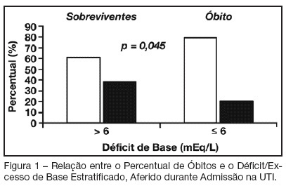
Views0See moreBACKGROUND AND OBJECTIVES: Base deficit is considered an indicator of tissue injury, shock and resuscitation. The objective of this study was to establish an association between base deficit obtained on the admission of patients in intensive care unit (ICU) and their prognosis. METHODS: A retrospective study with analysis of 110 patients admitted consecutively in the ICU, during the period of June to December 2006. RESULTS: There was a predominance of women, with age mean 54.2 ± 18.7 years old. Length of stay in ICU was 6.5 ± 7.4 days and the mean APACHE II score was 21 ± 8.1 points. The standardized mortality ratio was 0.715. Mortality was higher in patients with base deficit > 6 mEq/L (38.9%) than in those with base deficit < 6 mEq/L (20.6%); p < 0.05. Patients with early mortality had lower base deficit (7.75 ± 8.33 mEq/L) than survivors (3.17 ± 5.43 mEq/L); p < 0.05. Patients with permanence in ICU until 7 days and patients that stayed in this unit for more than 7 days had similar base deficit. CONCLUSIONS: Base deficit had been associated with early mortality during ICU internment. Base deficit > 6 mEq/L is a marker of significant mortality.
Views0Abstract
Base deficit at intensive care unit admission: an early mortality indicator
Rev Bras Ter Intensiva. 2007;19(4):434-436
DOI 10.1590/S0103-507X2007000400005
Views0See moreBACKGROUND AND OBJECTIVES: Base deficit is considered an indicator of tissue injury, shock and resuscitation. The objective of this study was to establish an association between base deficit obtained on the admission of patients in intensive care unit (ICU) and their prognosis. METHODS: A retrospective study with analysis of 110 patients admitted consecutively in the ICU, during the period of June to December 2006. RESULTS: There was a predominance of women, with age mean 54.2 ± 18.7 years old. Length of stay in ICU was 6.5 ± 7.4 days and the mean APACHE II score was 21 ± 8.1 points. The standardized mortality ratio was 0.715. Mortality was higher in patients with base deficit > 6 mEq/L (38.9%) than in those with base deficit < 6 mEq/L (20.6%); p < 0.05. Patients with early mortality had lower base deficit (7.75 ± 8.33 mEq/L) than survivors (3.17 ± 5.43 mEq/L); p < 0.05. Patients with permanence in ICU until 7 days and patients that stayed in this unit for more than 7 days had similar base deficit. CONCLUSIONS: Base deficit had been associated with early mortality during ICU internment. Base deficit > 6 mEq/L is a marker of significant mortality.
-
Partitioning evolutive standard base excess determinants in septic shock patients
Rev Bras Ter Intensiva. 2007;19(4):437-443
Abstract
Partitioning evolutive standard base excess determinants in septic shock patients
Rev Bras Ter Intensiva. 2007;19(4):437-443
DOI 10.1590/S0103-507X2007000400006
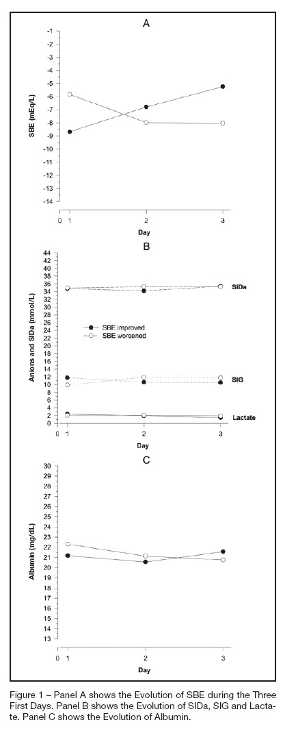
Views0See moreBACKGROUND AND OBJECTIVES: The amount of metabolic acidosis measured through the standard base excess (SBE) has been shown to be an outcome marker and its improvement has been associated with better survival. We studied the mechanism of standard base excess variation in the first three days of intensive care unit (ICU) stay through the evaluation of independent variables of physico-chemical approach. METHODS: Data were retrieved from our prospective collected data base from patients with diagnosis of septic shock, daily up to the third day after the ICU admission. Single correlations between SBE and independent variables were performed as well as a mathematical multilinear model was built to disclose the SBE variation determinants. RESULTS: We have shown that in septic shock patients the standard base excess variation during the first three days of ICU stay is weakly correlated to strong ion gap (SIG), lactate, creatinin and PaCO2 when individually analyzed. Analyzing concomitantly those independent variables, we built a mathematical model with a stepwise multilinear regression composed by apparent strong ion difference (SIDa), SIG, PaCO2, albumin and diuresis that resulted in a R² coefficient of 0.866 to determine SBE variation. CONCLUSIONS: Variations of metabolic acidosis measured through the standard base excess in septic shock patients when analyzed until the third day after intensive care unit admission, is resultant of interaction of several independent determinants as PaCO2, diuresis, SIG, SIDa and albumin.
Views0Abstract
Partitioning evolutive standard base excess determinants in septic shock patients
Rev Bras Ter Intensiva. 2007;19(4):437-443
DOI 10.1590/S0103-507X2007000400006
Views0See moreBACKGROUND AND OBJECTIVES: The amount of metabolic acidosis measured through the standard base excess (SBE) has been shown to be an outcome marker and its improvement has been associated with better survival. We studied the mechanism of standard base excess variation in the first three days of intensive care unit (ICU) stay through the evaluation of independent variables of physico-chemical approach. METHODS: Data were retrieved from our prospective collected data base from patients with diagnosis of septic shock, daily up to the third day after the ICU admission. Single correlations between SBE and independent variables were performed as well as a mathematical multilinear model was built to disclose the SBE variation determinants. RESULTS: We have shown that in septic shock patients the standard base excess variation during the first three days of ICU stay is weakly correlated to strong ion gap (SIG), lactate, creatinin and PaCO2 when individually analyzed. Analyzing concomitantly those independent variables, we built a mathematical model with a stepwise multilinear regression composed by apparent strong ion difference (SIDa), SIG, PaCO2, albumin and diuresis that resulted in a R² coefficient of 0.866 to determine SBE variation. CONCLUSIONS: Variations of metabolic acidosis measured through the standard base excess in septic shock patients when analyzed until the third day after intensive care unit admission, is resultant of interaction of several independent determinants as PaCO2, diuresis, SIG, SIDa and albumin.
-
A comparative study between early and late tracheostomy in patients ongoing mechanical ventilation
Rev Bras Ter Intensiva. 2007;19(4):444-449
Abstract
A comparative study between early and late tracheostomy in patients ongoing mechanical ventilation
Rev Bras Ter Intensiva. 2007;19(4):444-449
DOI 10.1590/S0103-507X2007000400007
Views0See moreBACKGROUND AND OBJECTIVES: To assess the incidence of tracheostomy in patients submitted to mechanic ventilation (MV); to compare the length of stay (LOS), duration of MV, APACHE II and mortality among patients submitted to tracheostomy, according to the moment of its application (early or late). METHODS: A retrospective observation study type cohort was done from April thru October 2005 including 190 patients at the adult intensive care unit (ICU) of Hospital Estadual do Grajaú. RESULTS: Among the 190 patients, 32 (16.84%) were submitted to tracheostomy with a longer LOS (30.16 days) as compared to those that were not (p < 0.001). The mean time of the tracheostomy procedure was 13.5 days from the starting of the MV. It is known that there still is no guidance for defining an ideal period for the operation. On this study, the prevalence of early tracheostomy (<13 days) was 46.87% (n = 15) and the late tracheostomy (> 13 days) was 53.13% (n = 17). In a meaningful way, the patients with early tracheostomy obtained APACHE II superior to those with late tracheostomy (18.2 versus 13.47), however there was no difference regarding the mortality rate. There was no difference regarding the time of ICU LOS (28.9 versus 31.28 days) and the MV time (29.73 versus 32.23 days) for both groups. CONCLUSIONS: The incidence of tracheostomy was high, being associated to a smaller ICU mortality but with a longer LOS and more complications. There was no significant difference regarding the destination of the patients when submitted to early or late tracheostomy.
Views0Abstract
A comparative study between early and late tracheostomy in patients ongoing mechanical ventilation
Rev Bras Ter Intensiva. 2007;19(4):444-449
DOI 10.1590/S0103-507X2007000400007
Views0See moreBACKGROUND AND OBJECTIVES: To assess the incidence of tracheostomy in patients submitted to mechanic ventilation (MV); to compare the length of stay (LOS), duration of MV, APACHE II and mortality among patients submitted to tracheostomy, according to the moment of its application (early or late). METHODS: A retrospective observation study type cohort was done from April thru October 2005 including 190 patients at the adult intensive care unit (ICU) of Hospital Estadual do Grajaú. RESULTS: Among the 190 patients, 32 (16.84%) were submitted to tracheostomy with a longer LOS (30.16 days) as compared to those that were not (p < 0.001). The mean time of the tracheostomy procedure was 13.5 days from the starting of the MV. It is known that there still is no guidance for defining an ideal period for the operation. On this study, the prevalence of early tracheostomy (<13 days) was 46.87% (n = 15) and the late tracheostomy (> 13 days) was 53.13% (n = 17). In a meaningful way, the patients with early tracheostomy obtained APACHE II superior to those with late tracheostomy (18.2 versus 13.47), however there was no difference regarding the mortality rate. There was no difference regarding the time of ICU LOS (28.9 versus 31.28 days) and the MV time (29.73 versus 32.23 days) for both groups. CONCLUSIONS: The incidence of tracheostomy was high, being associated to a smaller ICU mortality but with a longer LOS and more complications. There was no significant difference regarding the destination of the patients when submitted to early or late tracheostomy.
-
Central of mechanical fan: organization, safety and quality
Rev Bras Ter Intensiva. 2007;19(4):450-455
Abstract
Central of mechanical fan: organization, safety and quality
Rev Bras Ter Intensiva. 2007;19(4):450-455
DOI 10.1590/S0103-507X2007000400008
Views0See moreBACKGROUND AND OBJECTIVES: The headquarters of mechanical fans is the unit of the hospital with purpose of organizing ventilation resources promoting control and preventive maintenance and organizational of these equipments. The objective of this study was to elaborate a proposal of implantation of a headquarters of mechanical fans in an academical hospital, subsidized by the identification of the male nurse’s scientific technical knowledge on the theme ventilation mechanics and for the detection of problems originating from of the decentralized administration of the fans. METHODS: It is treated of exploratory descriptive study with quantitative approach, accomplished with 13 male nurses of ICU. The information was collected through structured interviews and submitted the descriptive analysis of the content. RESULTS: The results reveal that the male nurses possess several doubts, fact evidenced by 100% of the interviewees that mentioned the need of training courses gone back to the nursing attendance to the patient in ventilation mechanics. The situations described by the male nurses in the daily they demonstrate that the decentralization of the administration of the mechanical fans is shown ineffective as the organization, safety and quality. The proposal of implantation of a headquarters of fans appears for improvements in the attendance, in the formation of human resources and in the production of the knowledge. CONCLUSÕES: It is ended that the current profile can be changed through the breaking of institutional paradigms and of the institution of innovative practices that you/they will reinforce the purpose of a hospital of great load gone back to teaching, he/she researches and extension.
Views0Abstract
Central of mechanical fan: organization, safety and quality
Rev Bras Ter Intensiva. 2007;19(4):450-455
DOI 10.1590/S0103-507X2007000400008
Views0See moreBACKGROUND AND OBJECTIVES: The headquarters of mechanical fans is the unit of the hospital with purpose of organizing ventilation resources promoting control and preventive maintenance and organizational of these equipments. The objective of this study was to elaborate a proposal of implantation of a headquarters of mechanical fans in an academical hospital, subsidized by the identification of the male nurse’s scientific technical knowledge on the theme ventilation mechanics and for the detection of problems originating from of the decentralized administration of the fans. METHODS: It is treated of exploratory descriptive study with quantitative approach, accomplished with 13 male nurses of ICU. The information was collected through structured interviews and submitted the descriptive analysis of the content. RESULTS: The results reveal that the male nurses possess several doubts, fact evidenced by 100% of the interviewees that mentioned the need of training courses gone back to the nursing attendance to the patient in ventilation mechanics. The situations described by the male nurses in the daily they demonstrate that the decentralization of the administration of the mechanical fans is shown ineffective as the organization, safety and quality. The proposal of implantation of a headquarters of fans appears for improvements in the attendance, in the formation of human resources and in the production of the knowledge. CONCLUSÕES: It is ended that the current profile can be changed through the breaking of institutional paradigms and of the institution of innovative practices that you/they will reinforce the purpose of a hospital of great load gone back to teaching, he/she researches and extension.
-
Intensive care medicine on medical undergraduation: student’s perspective
Rev Bras Ter Intensiva. 2007;19(4):456-462
Abstract
Intensive care medicine on medical undergraduation: student’s perspective
Rev Bras Ter Intensiva. 2007;19(4):456-462
DOI 10.1590/S0103-507X2007000400009
Views1See moreBACKGROUND AND OBJECTIVES: There are deficiencies on Intensive Medicine (IM) teaching in most of medical undergraduate schools. Those deficiencies may imply damages on their clinical competence. The objective of this study was to analyze current status of IM teaching and the medical undergraduate student interest in this speciality. METHODS: A cross-sectional study was performed in 2005. We applied a self-reported questionnaire to enrolled students between the sixth and the last semesters of two medical schools from Salvador-Bahia. The questionnaire contained questions about students’ interest and knowledge on IM, and opinion on IM teaching in their schools. RESULTS: We studied 570 students. Most of them (57.5%) had never realized a clerkship in intensive care unit (ICU) despite classifying its usefulness as high (mean of 4.14 ± 1.05, in a scale from 1 to 5). IM interest was high or very high in 53.7% of sample. Almost all students (97%) thought that IM topics should be more explored at their curriculum. Only 42.1% reported to be able to assess a critical care patient and this assurance was higher among students with previous clerkship in ICU (p < 0.001). Shock, cardiopulmonary resuscitation and sepsis were the most interesting topics in ICU for students' opinion. CONCLUSIONS: This study revealed a high interest in IM among medical undergraduate students. However, most had never practice a clerkship in ICU, demonstrating to be an important factor on undergraduate student performance faced to a critical care patient.
Views1Abstract
Intensive care medicine on medical undergraduation: student’s perspective
Rev Bras Ter Intensiva. 2007;19(4):456-462
DOI 10.1590/S0103-507X2007000400009
Views1See moreBACKGROUND AND OBJECTIVES: There are deficiencies on Intensive Medicine (IM) teaching in most of medical undergraduate schools. Those deficiencies may imply damages on their clinical competence. The objective of this study was to analyze current status of IM teaching and the medical undergraduate student interest in this speciality. METHODS: A cross-sectional study was performed in 2005. We applied a self-reported questionnaire to enrolled students between the sixth and the last semesters of two medical schools from Salvador-Bahia. The questionnaire contained questions about students’ interest and knowledge on IM, and opinion on IM teaching in their schools. RESULTS: We studied 570 students. Most of them (57.5%) had never realized a clerkship in intensive care unit (ICU) despite classifying its usefulness as high (mean of 4.14 ± 1.05, in a scale from 1 to 5). IM interest was high or very high in 53.7% of sample. Almost all students (97%) thought that IM topics should be more explored at their curriculum. Only 42.1% reported to be able to assess a critical care patient and this assurance was higher among students with previous clerkship in ICU (p < 0.001). Shock, cardiopulmonary resuscitation and sepsis were the most interesting topics in ICU for students' opinion. CONCLUSIONS: This study revealed a high interest in IM among medical undergraduate students. However, most had never practice a clerkship in ICU, demonstrating to be an important factor on undergraduate student performance faced to a critical care patient.
-
Disseminated strongyloidiasis: diagnosis and treatment
Rev Bras Ter Intensiva. 2007;19(4):463-468
Abstract
Disseminated strongyloidiasis: diagnosis and treatment
Rev Bras Ter Intensiva. 2007;19(4):463-468
DOI 10.1590/S0103-507X2007000400010
Views0See moreBACKGROUND AND OBJECTIVES: Disseminated strongyloidiasis is a clinical form of presentation associated with states of severe immunosuppression, as in AIDS, hematological malignancies and in treatment for immunosuppression (especially with high doses of corticosteroids). It usually mimics severe sepsis and still brings a significant challenge related to the diagnosis and treatment. Therefore exceedingly high mortality rates remain unchanged in the past decades. Initially, the diagnosis depends on the clinical suspicion and on the identification of the larva in an organic fluids or tissues. The cutaneous involvement, albeit rare, is typical and can provide an important clue for the diagnostic hypothesis. The emergence of ivermectin for oral use changed significantly the treatment for strongyloidiasis; however, there are still shortcomings for the utilization in critically ill patients. Shock, ileus and hypoperfusion states are associated with difficulties in the absorption that result in erratic systemic levels. Reports of good results with parenteral administration of ivermectin raised the prospect that this therapeutic modality be more effective. However, questions about dosage and safety remain unanswered. The aim of the present article is to review the medical literature on the clinical aspects of disseminated strongyloidiasis. CONTENTS: A systematic review of the literature was performed by searching the PubMed database within the last 30 years. Search terms were: disseminated strongyloidiasis, strongyloides and hyperinfection e ivermectin. CONCLUSIONS: The article highlights the diagnostic and therapeutic aspects emphasizing the importance of the clinical suspicion for the institution of appropriated therapy.
Views0Abstract
Disseminated strongyloidiasis: diagnosis and treatment
Rev Bras Ter Intensiva. 2007;19(4):463-468
DOI 10.1590/S0103-507X2007000400010
Views0See moreBACKGROUND AND OBJECTIVES: Disseminated strongyloidiasis is a clinical form of presentation associated with states of severe immunosuppression, as in AIDS, hematological malignancies and in treatment for immunosuppression (especially with high doses of corticosteroids). It usually mimics severe sepsis and still brings a significant challenge related to the diagnosis and treatment. Therefore exceedingly high mortality rates remain unchanged in the past decades. Initially, the diagnosis depends on the clinical suspicion and on the identification of the larva in an organic fluids or tissues. The cutaneous involvement, albeit rare, is typical and can provide an important clue for the diagnostic hypothesis. The emergence of ivermectin for oral use changed significantly the treatment for strongyloidiasis; however, there are still shortcomings for the utilization in critically ill patients. Shock, ileus and hypoperfusion states are associated with difficulties in the absorption that result in erratic systemic levels. Reports of good results with parenteral administration of ivermectin raised the prospect that this therapeutic modality be more effective. However, questions about dosage and safety remain unanswered. The aim of the present article is to review the medical literature on the clinical aspects of disseminated strongyloidiasis. CONTENTS: A systematic review of the literature was performed by searching the PubMed database within the last 30 years. Search terms were: disseminated strongyloidiasis, strongyloides and hyperinfection e ivermectin. CONCLUSIONS: The article highlights the diagnostic and therapeutic aspects emphasizing the importance of the clinical suspicion for the institution of appropriated therapy.
-
Understanding the mechanisms of ventilator-induced lung injury
Rev Bras Ter Intensiva. 2007;19(4):469-474
Abstract
Understanding the mechanisms of ventilator-induced lung injury
Rev Bras Ter Intensiva. 2007;19(4):469-474
DOI 10.1590/S0103-507X2007000400011
Views0See moreBACKGROUND AND OBJECTIVES: Mechanical ventilation is considered a basic element of life support in the intensive care unit and is essential for patients with acute lung injury (ALI) and acute respiratory distress syndrome (ARDS). Experimental studies have demonstrated that mechanical ventilation with high volumes and/or high pressures can exacerbate (VALI) or induce lung injury (VILI) with histological aspect similar to ALI/ARDS. CONTENTS: This systematic review included the literature on MedLine and SciElo database published in the last 20 years. In this review, we will highlight the most recent data on the mechanisms of VILI. The main mechanisms of VILI are: volutrauma caused by overinflation and uneven expansion of the lungs due to high ventilation pressures or volumes; aletectrauma induced by shear forces generated during cyclic closure and reopening of terminal airways; and biotrauma where the injury resulted from the release inflammatory mediators due to physical stresses associated with mechanical ventilation. CONCLUSIONS: It is fundamental to understand the mechanisms related to volutrauma, atelectrauma, and biotrauma to avoid ventilator-associated lung injury.
Views0Abstract
Understanding the mechanisms of ventilator-induced lung injury
Rev Bras Ter Intensiva. 2007;19(4):469-474
DOI 10.1590/S0103-507X2007000400011
Views0See moreBACKGROUND AND OBJECTIVES: Mechanical ventilation is considered a basic element of life support in the intensive care unit and is essential for patients with acute lung injury (ALI) and acute respiratory distress syndrome (ARDS). Experimental studies have demonstrated that mechanical ventilation with high volumes and/or high pressures can exacerbate (VALI) or induce lung injury (VILI) with histological aspect similar to ALI/ARDS. CONTENTS: This systematic review included the literature on MedLine and SciElo database published in the last 20 years. In this review, we will highlight the most recent data on the mechanisms of VILI. The main mechanisms of VILI are: volutrauma caused by overinflation and uneven expansion of the lungs due to high ventilation pressures or volumes; aletectrauma induced by shear forces generated during cyclic closure and reopening of terminal airways; and biotrauma where the injury resulted from the release inflammatory mediators due to physical stresses associated with mechanical ventilation. CONCLUSIONS: It is fundamental to understand the mechanisms related to volutrauma, atelectrauma, and biotrauma to avoid ventilator-associated lung injury.
-
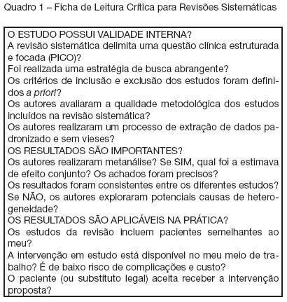
How to critically assess systematic reviews and meta-analyses?
Rev Bras Ter Intensiva. 2007;19(4):475-480
Abstract
How to critically assess systematic reviews and meta-analyses?
Rev Bras Ter Intensiva. 2007;19(4):475-480
DOI 10.1590/S0103-507X2007000400012
Views0See moreBACKGROUND AND OBJECTIVES: Systematic reviews are important knowledge generating tools to help the decision making process in the Critical Care Unit. This narrative aims to describe the important elements used to critically appraise intensive care-related systematic reviews. CONTENTS: When critically assessing systematic reviews, one should pay particular attention to the importance and appropriateness of the research question, the search strategy, the inclusion criteria and methodological quality of the studies included, and the methods of data extraction. In addition, a relevant systematic review must have consistent data (in case of a meta-analysis) or the cause of the heterogeneity must have been adequately explored, and results must be applicable in critical patients. CONCLUSIONS: To apply correctly the available scientific evidence, one should critically assess data quality of systematic reviews, selecting the relevant information to manage the critically ill patient.
Views0Abstract
How to critically assess systematic reviews and meta-analyses?
Rev Bras Ter Intensiva. 2007;19(4):475-480
DOI 10.1590/S0103-507X2007000400012
Views0See moreBACKGROUND AND OBJECTIVES: Systematic reviews are important knowledge generating tools to help the decision making process in the Critical Care Unit. This narrative aims to describe the important elements used to critically appraise intensive care-related systematic reviews. CONTENTS: When critically assessing systematic reviews, one should pay particular attention to the importance and appropriateness of the research question, the search strategy, the inclusion criteria and methodological quality of the studies included, and the methods of data extraction. In addition, a relevant systematic review must have consistent data (in case of a meta-analysis) or the cause of the heterogeneity must have been adequately explored, and results must be applicable in critical patients. CONCLUSIONS: To apply correctly the available scientific evidence, one should critically assess data quality of systematic reviews, selecting the relevant information to manage the critically ill patient.
-
Caring for the families of terminally ill patients in the intensive care unit
Rev Bras Ter Intensiva. 2007;19(4):481-484
Abstract
Caring for the families of terminally ill patients in the intensive care unit
Rev Bras Ter Intensiva. 2007;19(4):481-484
DOI 10.1590/S0103-507X2007000400013
Views0See moreBACKGROUND AND OBJECTIVES: Caring for the families is one of the major tasks of the global care of patients admitted to the intensive care unit (ICU). In the context of a terminally ill patient or a patient in whom the recovery from the acute illness is unlikely, dealing with and caring for their family members becomes even more important as the patient will not be awake in most of situations. Family members have specific needs and present with high incidence of symptoms of stress, depression, anxiety and related disorders during the ICU of their beloved one, which can even persist late after the patient’s death. CONTENTS: Review of selected studies on the care of family members of patients at the end-of-life admitted to the ICU published at the PubMed database during the last 20 years. CONCLUSIONS: Recent literature is plenty of evidence that strategies directed to care of family members, such as improvement of the communication process, prevention of conflicts, and spiritual care, can improve satisfaction and perception of quality on the care of patients at the end-of-life in the ICU.
Views0Abstract
Caring for the families of terminally ill patients in the intensive care unit
Rev Bras Ter Intensiva. 2007;19(4):481-484
DOI 10.1590/S0103-507X2007000400013
Views0See moreBACKGROUND AND OBJECTIVES: Caring for the families is one of the major tasks of the global care of patients admitted to the intensive care unit (ICU). In the context of a terminally ill patient or a patient in whom the recovery from the acute illness is unlikely, dealing with and caring for their family members becomes even more important as the patient will not be awake in most of situations. Family members have specific needs and present with high incidence of symptoms of stress, depression, anxiety and related disorders during the ICU of their beloved one, which can even persist late after the patient’s death. CONTENTS: Review of selected studies on the care of family members of patients at the end-of-life admitted to the ICU published at the PubMed database during the last 20 years. CONCLUSIONS: Recent literature is plenty of evidence that strategies directed to care of family members, such as improvement of the communication process, prevention of conflicts, and spiritual care, can improve satisfaction and perception of quality on the care of patients at the end-of-life in the ICU.
-
How to improve the communication and to prevent the conflicts at terminality situations in Intensive Care Unit
Rev Bras Ter Intensiva. 2007;19(4):485-489
Abstract
How to improve the communication and to prevent the conflicts at terminality situations in Intensive Care Unit
Rev Bras Ter Intensiva. 2007;19(4):485-489
DOI 10.1590/S0103-507X2007000400014
Views0See moreBACKGROUND AND OBJECTIVES: The suffering with the death and the prolonged time of patient’s admission in a intensive care unit (ICU) are factors that leads to necessity the best communication with the personal that works in ICU, patients and their family, and its justify this work, whose objective is discuss this subject. CONTENTS: The professional experience of the author was used in this issue and the articles written during the last five years about death, communication and ICU were reviewed by means of MedLine, Up to Date, Google and Brazilian Journal of Intensive Therapy. CONCLUSIONS: It was concluded that the physician, the patient together with her/his family, and the multiprofessional staff of the ICU is one of the main factors that interferes with the process of satisfying both the patient and the ones who work on such unities. For adequate information, the physician must be conscious about therapeutic limits, and must learn how to treat the patient during the process of dying. In this way, the physician will be apt to talk about death. The ideal situation would be that the professional, responsible to give the news, should be experience, from the technical point of view as well as ethical, and should be the same person, as always as possible, when necessary. The patient mostly little be able to influence in her/his process of dying, but if communication is possible, it be simple, honest and humane. The patient’s family members have the right of being together with the one who they love, and of being steadily informed about the real situation. All the members in the process must know the truth and the chosen therapeutic orientation to be taken. Communication should be done in a quiet and prived place.
Views0Abstract
How to improve the communication and to prevent the conflicts at terminality situations in Intensive Care Unit
Rev Bras Ter Intensiva. 2007;19(4):485-489
DOI 10.1590/S0103-507X2007000400014
Views0See moreBACKGROUND AND OBJECTIVES: The suffering with the death and the prolonged time of patient’s admission in a intensive care unit (ICU) are factors that leads to necessity the best communication with the personal that works in ICU, patients and their family, and its justify this work, whose objective is discuss this subject. CONTENTS: The professional experience of the author was used in this issue and the articles written during the last five years about death, communication and ICU were reviewed by means of MedLine, Up to Date, Google and Brazilian Journal of Intensive Therapy. CONCLUSIONS: It was concluded that the physician, the patient together with her/his family, and the multiprofessional staff of the ICU is one of the main factors that interferes with the process of satisfying both the patient and the ones who work on such unities. For adequate information, the physician must be conscious about therapeutic limits, and must learn how to treat the patient during the process of dying. In this way, the physician will be apt to talk about death. The ideal situation would be that the professional, responsible to give the news, should be experience, from the technical point of view as well as ethical, and should be the same person, as always as possible, when necessary. The patient mostly little be able to influence in her/his process of dying, but if communication is possible, it be simple, honest and humane. The patient’s family members have the right of being together with the one who they love, and of being steadily informed about the real situation. All the members in the process must know the truth and the chosen therapeutic orientation to be taken. Communication should be done in a quiet and prived place.
-
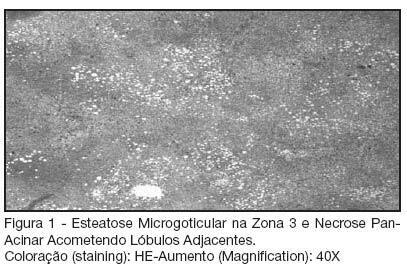
Liver ischemic necrosis and diabetes mellitus: case report
Rev Bras Ter Intensiva. 2007;19(4):490-493
Abstract
Liver ischemic necrosis and diabetes mellitus: case report
Rev Bras Ter Intensiva. 2007;19(4):490-493
DOI 10.1590/S0103-507X2007000400015
Views1See moreBACKGROUND AND OBJECTIVES: Hepatic infarction is characterized by parenchyma ischemic necrosis involving at least two acinis. It is extremely uncommon due to the arterial and portal venous blood supply. We report a case of a patient not know to have diabetes who developed massive areas of ischemic infarcts of the liver after episode of acutely diabetes decompensated. CASE REPORT: A 67 year-old hypertensive female who has been presenting, for the last 10 days, polydipsia, high urinary volume, visual and gait impairment, nausea and vomiting was admitted to the emergency room (ER). During the physical examination it was observed dehydration, skin discoloration, peripheral cyanosis, hypothermia, tachycardia, hypotension and mild diffuse abdominal pain. Admissional laboratory exams demonstrated total leukocytes: 16.800, Cr: 3.7, Ur: 167, Na: 133, K: 6.9, glucose: 561; arterial gasometry (O2 catheter: 2 L/min): pH: 6.93, pCO2: 12.1, pO2: 107, B.E.: -28.8, HCO3: 2.4, Sat 91.3%, lactato: 79; urinalysis: pH: 6; leukocytes: 13; density: 1015; erythrocytes: 19; protein: ++; glucose: +++; bilirubin: negative; ketonic bodies: + denote ketonemia. EKG: sharp T wave, right branch block. Patient was treated with intravenous insulin, hydration, sodium bicarbonate and ceftriaxone. After initial treatment, the laboratory exams showed Cr: 2.2, Ur: 122, Na: 162, K: 4.3, Ca: 6.4, glucose: 504, pH: 7.01, HCO3: 7.1, B.E.: -22. One day after admission the patient presented with important abdominal pain and peritoneal irritation, followed by difficulty for talking and somnolence; routine laboratory exams showed arterial gasometry: pH: 7.4, pCO2: 31, pO2: 68, BE: -4.4, HCO3: 19, SatO2: 93.5%; Ur: 95,Cr: 1.4, albumin: 2.4, Ca: 0.95, Na: 166, K:4, bilirubin: 0.5, bilirubin D/I: 0.2/0.3, Amylase: 1157, Gamma-GT: 56, AST 7.210, ALT: 2.470, SR (sedimentation rate): 15, Lipase: 84. Abdominal ultrasound was unremarkable. Patient respiratory function and conscience level worsened, requiring intubation. Despite all resuscitation efforts, she died. Necropsy showed multiple ischemic infarcts of the liver with vascular thrombosis, splenic infarcts, generalized visceral congestion and atherosclerosis of aorta and its branches. Pancreas was normal. CONCLUSIONS: The mechanisms of hepatic and splenic infarctions in this case were unclear. The following factors may have contributed to necrosis: vomiting and fever should be considered to induce dehydration and hypotension, which further decreased portal and hepatic arterial inflows; elevated level of catecholamine in hyperglycemic states might induce vasoconstriction effects; widespread atherosclerosis is commonly seen in diabetic and hypertensive patients. This case underlies the importance of searching for hepatic necrosis or infarction in any diabetic patient with elevated liver enzymes. Anticoagulation therapy should be instituted promptly upon recognition of vascular thromboses.
Views1Abstract
Liver ischemic necrosis and diabetes mellitus: case report
Rev Bras Ter Intensiva. 2007;19(4):490-493
DOI 10.1590/S0103-507X2007000400015
Views1See moreBACKGROUND AND OBJECTIVES: Hepatic infarction is characterized by parenchyma ischemic necrosis involving at least two acinis. It is extremely uncommon due to the arterial and portal venous blood supply. We report a case of a patient not know to have diabetes who developed massive areas of ischemic infarcts of the liver after episode of acutely diabetes decompensated. CASE REPORT: A 67 year-old hypertensive female who has been presenting, for the last 10 days, polydipsia, high urinary volume, visual and gait impairment, nausea and vomiting was admitted to the emergency room (ER). During the physical examination it was observed dehydration, skin discoloration, peripheral cyanosis, hypothermia, tachycardia, hypotension and mild diffuse abdominal pain. Admissional laboratory exams demonstrated total leukocytes: 16.800, Cr: 3.7, Ur: 167, Na: 133, K: 6.9, glucose: 561; arterial gasometry (O2 catheter: 2 L/min): pH: 6.93, pCO2: 12.1, pO2: 107, B.E.: -28.8, HCO3: 2.4, Sat 91.3%, lactato: 79; urinalysis: pH: 6; leukocytes: 13; density: 1015; erythrocytes: 19; protein: ++; glucose: +++; bilirubin: negative; ketonic bodies: + denote ketonemia. EKG: sharp T wave, right branch block. Patient was treated with intravenous insulin, hydration, sodium bicarbonate and ceftriaxone. After initial treatment, the laboratory exams showed Cr: 2.2, Ur: 122, Na: 162, K: 4.3, Ca: 6.4, glucose: 504, pH: 7.01, HCO3: 7.1, B.E.: -22. One day after admission the patient presented with important abdominal pain and peritoneal irritation, followed by difficulty for talking and somnolence; routine laboratory exams showed arterial gasometry: pH: 7.4, pCO2: 31, pO2: 68, BE: -4.4, HCO3: 19, SatO2: 93.5%; Ur: 95,Cr: 1.4, albumin: 2.4, Ca: 0.95, Na: 166, K:4, bilirubin: 0.5, bilirubin D/I: 0.2/0.3, Amylase: 1157, Gamma-GT: 56, AST 7.210, ALT: 2.470, SR (sedimentation rate): 15, Lipase: 84. Abdominal ultrasound was unremarkable. Patient respiratory function and conscience level worsened, requiring intubation. Despite all resuscitation efforts, she died. Necropsy showed multiple ischemic infarcts of the liver with vascular thrombosis, splenic infarcts, generalized visceral congestion and atherosclerosis of aorta and its branches. Pancreas was normal. CONCLUSIONS: The mechanisms of hepatic and splenic infarctions in this case were unclear. The following factors may have contributed to necrosis: vomiting and fever should be considered to induce dehydration and hypotension, which further decreased portal and hepatic arterial inflows; elevated level of catecholamine in hyperglycemic states might induce vasoconstriction effects; widespread atherosclerosis is commonly seen in diabetic and hypertensive patients. This case underlies the importance of searching for hepatic necrosis or infarction in any diabetic patient with elevated liver enzymes. Anticoagulation therapy should be instituted promptly upon recognition of vascular thromboses.
-
Hantavirus pulmonary syndrome with multiple organ dysfunctions: case report
Rev Bras Ter Intensiva. 2007;19(4):494-498
Abstract
Hantavirus pulmonary syndrome with multiple organ dysfunctions: case report
Rev Bras Ter Intensiva. 2007;19(4):494-498
DOI 10.1590/S0103-507X2007000400016
Views0BACKGROUND AND OBJECTIVES: Hantavirus infection is a zoonose with worldwide distribution. The transmission is related to the intimal contact with rodents. It causes two syndromes: hemorrhagic fever with renal syndrome (HFRS), endemic in Asia and Europe and the Hantavirus pulmonary syndrome (HPS), found in the American continent, including Brazil, with high mortality rates. The aim of this study is to report a case of HPS with multiple organ failure, managed with early goal-directed therapy guided by flow and tissue perfusion parameters. CASE REPORT: A 36 year-old male had fever with progressive dispnea, severe hypoxia and acute respiratory failure. Diffuse interstitial alveolar infiltrates were seen in the chest X-Ray. He developed multiple organ dysfunction syndromes (pulmonary, renal, coagulation, cardiovascular and metabolic). Treatment and invasive hemodynamic monitoring with pulmonary artery catheter was early instituted. The most important laboratory findings were thrombocytopenia, elevated hematocrit and hemoglobin concentrations, elevated liver enzymes, elevated lactate dehydrogenase and a positive sorology for Hantavirus (ELISA IgM positive). Organ dysfunctions reverted to normal and he was discharged after 21 days in hospital. CONCLUSIONS: An early and adequate resuscitation with goal-directed therapy enabled the reversion of the multiple organ failure syndromes and a favorable outcome, despite the severity of the disease.
Keywords:HantavirusHantavirus Cardiopulmonary SyndromeHantavirus pulmonary syndromemultiple organ dysfunctionSee moreViews0Abstract
Hantavirus pulmonary syndrome with multiple organ dysfunctions: case report
Rev Bras Ter Intensiva. 2007;19(4):494-498
DOI 10.1590/S0103-507X2007000400016
Views0BACKGROUND AND OBJECTIVES: Hantavirus infection is a zoonose with worldwide distribution. The transmission is related to the intimal contact with rodents. It causes two syndromes: hemorrhagic fever with renal syndrome (HFRS), endemic in Asia and Europe and the Hantavirus pulmonary syndrome (HPS), found in the American continent, including Brazil, with high mortality rates. The aim of this study is to report a case of HPS with multiple organ failure, managed with early goal-directed therapy guided by flow and tissue perfusion parameters. CASE REPORT: A 36 year-old male had fever with progressive dispnea, severe hypoxia and acute respiratory failure. Diffuse interstitial alveolar infiltrates were seen in the chest X-Ray. He developed multiple organ dysfunction syndromes (pulmonary, renal, coagulation, cardiovascular and metabolic). Treatment and invasive hemodynamic monitoring with pulmonary artery catheter was early instituted. The most important laboratory findings were thrombocytopenia, elevated hematocrit and hemoglobin concentrations, elevated liver enzymes, elevated lactate dehydrogenase and a positive sorology for Hantavirus (ELISA IgM positive). Organ dysfunctions reverted to normal and he was discharged after 21 days in hospital. CONCLUSIONS: An early and adequate resuscitation with goal-directed therapy enabled the reversion of the multiple organ failure syndromes and a favorable outcome, despite the severity of the disease.
Keywords:HantavirusHantavirus Cardiopulmonary SyndromeHantavirus pulmonary syndromemultiple organ dysfunctionSee more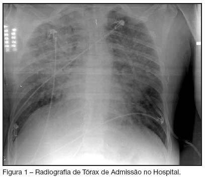
-
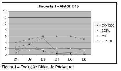
Macrophage migration inhibitory factor and interleukin-6 in crush syndrome: analogy with severity? Case reports
Rev Bras Ter Intensiva. 2007;19(4):499-503
Abstract
Macrophage migration inhibitory factor and interleukin-6 in crush syndrome: analogy with severity? Case reports
Rev Bras Ter Intensiva. 2007;19(4):499-503
DOI 10.1590/S0103-507X2007000400017
Views0See moreBACKGROUND AND OBJECTIVES: Macrophage migration inhibitory factor (MIF) is a multifunctional cytokine involved in a broad-spectrum pathological events relevant to the immune system. Interleukin-6 (IL-6) is a proinflammatory cytokine that plays an important role in the initial inflammatory response to trauma and the development of early and late multiple organ dysfunction syndrome (MODS). Crush syndrome has been described as the systemic manifestation of muscle cell damage resulting from pressing or crushing. There are few data about MIF and IL-6 in crush syndrome. The aim of this study was to report four cases of crush syndrome, measuring seric levels of MIF and IL-6 and its correlation with severity. CASES REPORTS: Four patients suffering from crush syndrome after an accident with an explosive artifact were enrolled in the study. APACHE II score was checked at admission. It was collected serum sample of these patients during six consecutive days. Serum MIF, IL-6 and creatine kinase (CK) were measured. Sepsis-related organ failure assessment (SOFA) score was evaluated concomitantly. Data were analyzed. CONCLUSIONS: The variations observed in the CK measures were followed by alterations in the cytokines’ level and at the SOFA score, suggesting interdependence between those factors. Other articles have already demonstrated similar results. Although the use of cytokines as biomarkers of severity in trauma is matter of interest, we need large studies with a higher number of patients to validate this observation.
Views0Abstract
Macrophage migration inhibitory factor and interleukin-6 in crush syndrome: analogy with severity? Case reports
Rev Bras Ter Intensiva. 2007;19(4):499-503
DOI 10.1590/S0103-507X2007000400017
Views0See moreBACKGROUND AND OBJECTIVES: Macrophage migration inhibitory factor (MIF) is a multifunctional cytokine involved in a broad-spectrum pathological events relevant to the immune system. Interleukin-6 (IL-6) is a proinflammatory cytokine that plays an important role in the initial inflammatory response to trauma and the development of early and late multiple organ dysfunction syndrome (MODS). Crush syndrome has been described as the systemic manifestation of muscle cell damage resulting from pressing or crushing. There are few data about MIF and IL-6 in crush syndrome. The aim of this study was to report four cases of crush syndrome, measuring seric levels of MIF and IL-6 and its correlation with severity. CASES REPORTS: Four patients suffering from crush syndrome after an accident with an explosive artifact were enrolled in the study. APACHE II score was checked at admission. It was collected serum sample of these patients during six consecutive days. Serum MIF, IL-6 and creatine kinase (CK) were measured. Sepsis-related organ failure assessment (SOFA) score was evaluated concomitantly. Data were analyzed. CONCLUSIONS: The variations observed in the CK measures were followed by alterations in the cytokines’ level and at the SOFA score, suggesting interdependence between those factors. Other articles have already demonstrated similar results. Although the use of cytokines as biomarkers of severity in trauma is matter of interest, we need large studies with a higher number of patients to validate this observation.
-
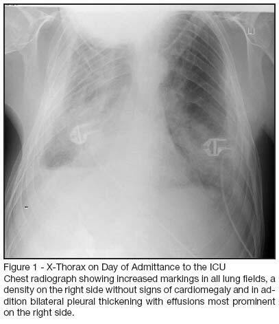
Failure to wean caused by cryptogenic fibrosing pleuritis and bilateral lung trapping: case report
Rev Bras Ter Intensiva. 2007;19(4):504-508
Abstract
Failure to wean caused by cryptogenic fibrosing pleuritis and bilateral lung trapping: case report
Rev Bras Ter Intensiva. 2007;19(4):504-508
DOI 10.1590/S0103-507X2007000400018
Views0See moreBACKGROUND AND OBJECTIVES: Cryptogenic fibrosing pleuritis is an extremely rare disease, which can affect both lungs from a very young age. The most common finding is severe lung restriction resulting in both hypoxemic and ventilatory failure. CASE REPORT: Male patient, 26 year old with acute deterioration of chronic respiratory failure. Following admission prolonged mechanical ventilation was necessary. An atypical clinical presentation made the diagnosis difficult, but eventually cryptogenic fibrosing pleuritis and lung fibrosis were established. CONCLUSIONS: The prognostic outcome of patients with the final diagnosis of cryptogenic fibrosing pleuritis is extremely poor, especially in an advanced phase of this disease. We recommend early treatment with corticosteroids or surgical pleural decortication.
Views0Abstract
Failure to wean caused by cryptogenic fibrosing pleuritis and bilateral lung trapping: case report
Rev Bras Ter Intensiva. 2007;19(4):504-508
DOI 10.1590/S0103-507X2007000400018
Views0See moreBACKGROUND AND OBJECTIVES: Cryptogenic fibrosing pleuritis is an extremely rare disease, which can affect both lungs from a very young age. The most common finding is severe lung restriction resulting in both hypoxemic and ventilatory failure. CASE REPORT: Male patient, 26 year old with acute deterioration of chronic respiratory failure. Following admission prolonged mechanical ventilation was necessary. An atypical clinical presentation made the diagnosis difficult, but eventually cryptogenic fibrosing pleuritis and lung fibrosis were established. CONCLUSIONS: The prognostic outcome of patients with the final diagnosis of cryptogenic fibrosing pleuritis is extremely poor, especially in an advanced phase of this disease. We recommend early treatment with corticosteroids or surgical pleural decortication.
Volume Articles - Critical Care Science (CCS)
